Msa Plate Staphylococcus Epidermidis

Pin On Micro
Mannitol Salt Agar Msa Test Scientist Cindy
Mannitol Utilisation Is Required For Protection Of Staphylococcus Aureus From Human Skin Antimicrobial Fatty Acids

S Aureus And E Faecalis Colonies Obtained By Culturing Them On Download Scientific Diagram

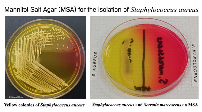
Yellow Colonies Of S Aureus On Mannitol Salt Agar Download Scientific Diagram

Emb Agar Only Gram Negative Bacteria Grow On Emb Agar Gram Positive Bacteria Are Inhibited By The Dyes Eosin And Methylene Blue Added To The Agar Based Ppt Video Online Download
Staphylococcus epidermidis is a gram-positive spherical bacterium that form irregular clusters.

Msa plate staphylococcus epidermidis. Mannitol Salt Agar (MSA) Plate. Mannitol Salt Agar (MSA) is used as a selective and differential medium for the isolation and identification of Staphylococcus aureus from clinical and non-clinical specimens. It encourages the growth of a group of certain bacteria while inhibiting the growth of others.
Mannitol Salt Agar, Methyl Red, Catalase, and Urea. Gram stain each of the organisms;. Day Three Individual Supplies share Blood Agar plates 1½ NA plates & antibiotic disk dispenser containing Novobiocin 1.
The observations after the incubation on the Mannitol Salt Agar concluded that the bacterium ferments mannitol. Subculture TSB, USP enrichemnt to MSA, USP, and incubate at 30-35 o C for 18-72 hours. An MSA plate with Micrococcus sp.
Staphylococcus is a genus of Gram +, nonspore-forming cocci belonging to the family Micrococcaceae that are often found as normal human microbiota of the skin and nasal cavity.There are five organisms to consider as potential human pathogens in this genus:. Aseptically remove lid of contact plate. In other words, Staphylococcus epidermis is considered "coagulase -" or "coagulase-negative".
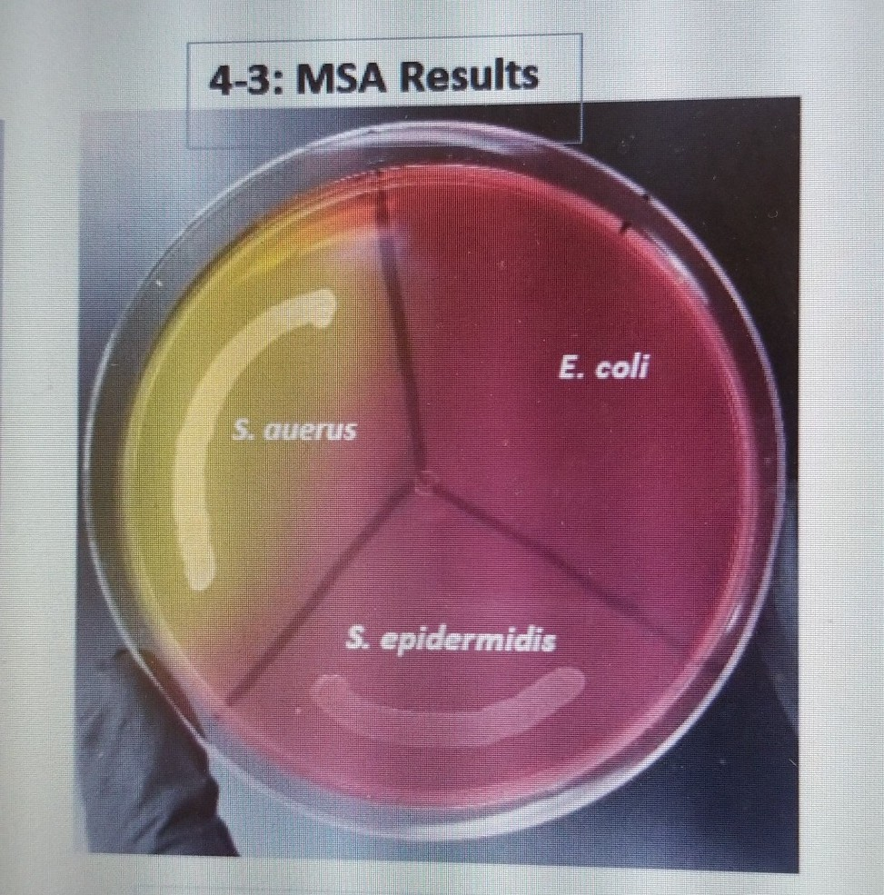
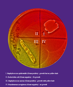
Staphylococcus epidermidis (Gram positive) - growth but no yellow halo. An MSA plate with Micrococcus sp. It is part of the normal human flora, typically the skin flora, and less commonly the mucosal flora.
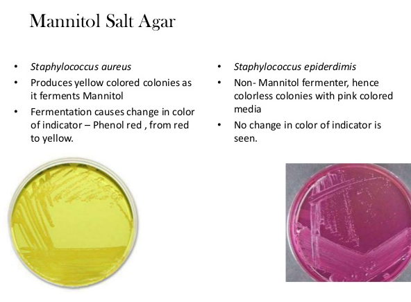
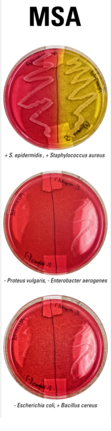
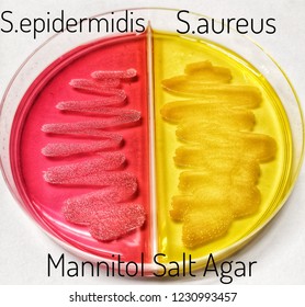
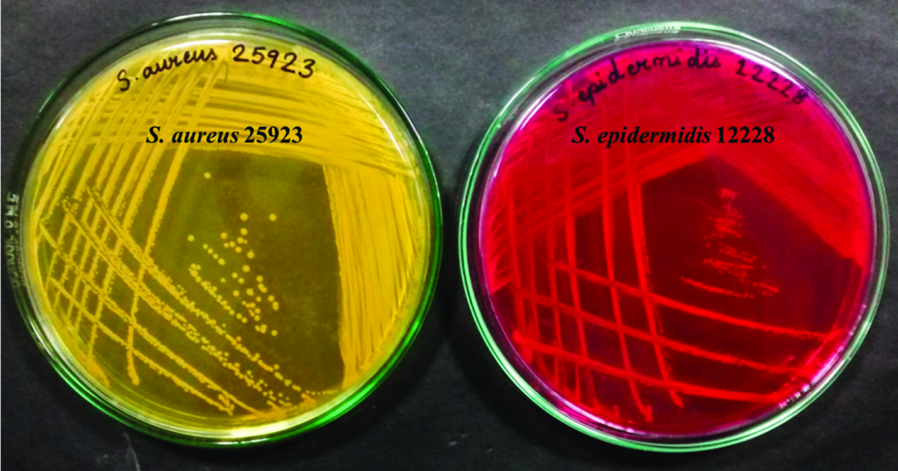
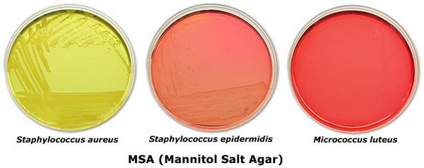
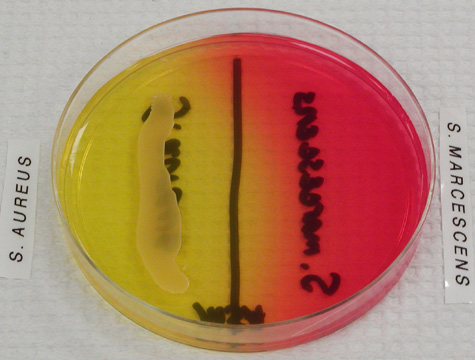
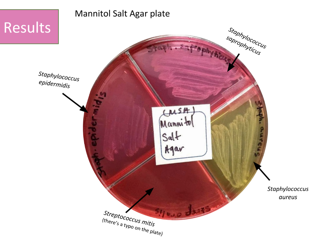
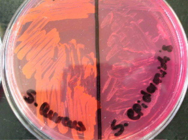
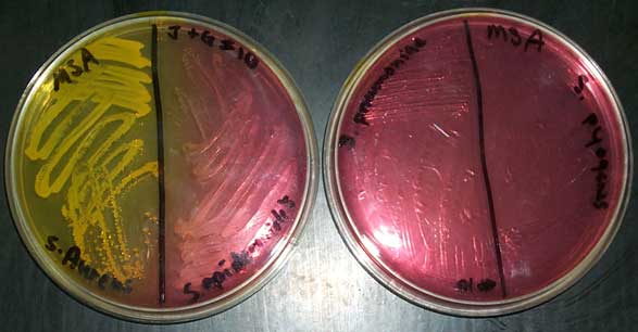
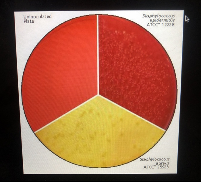
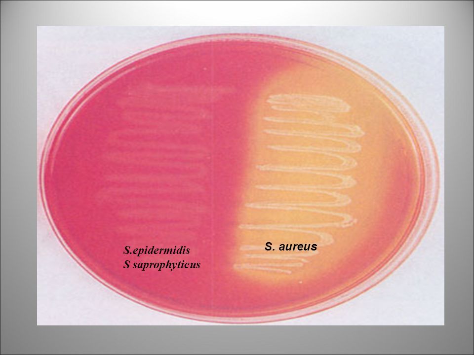
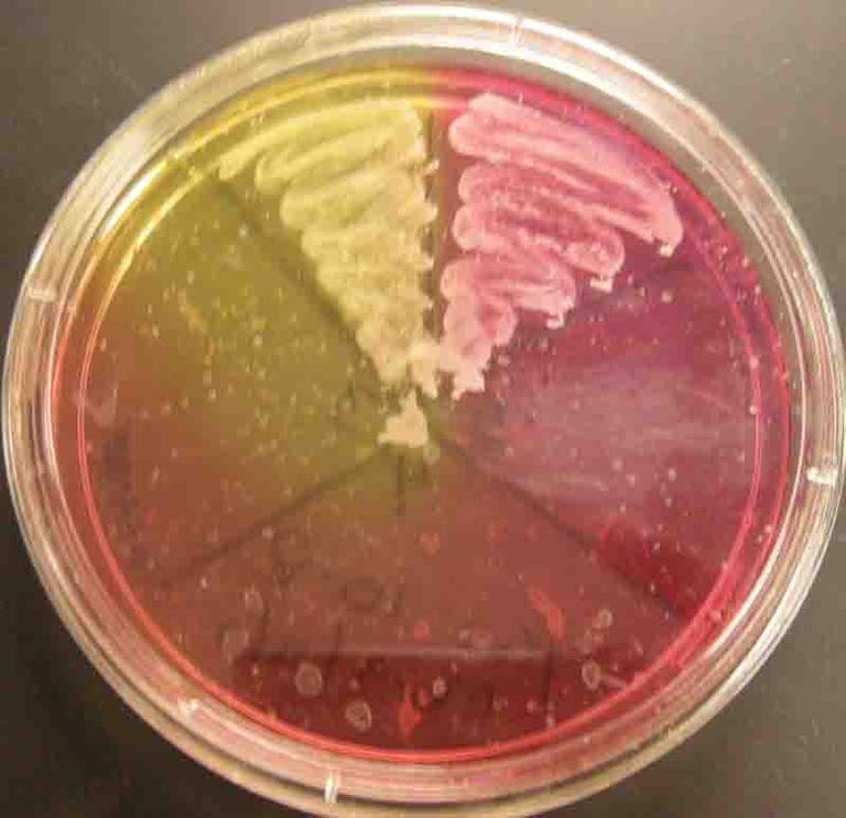
Staphylococcus aureus and Staphylococcus epidermidis on mannitol salt agar. Growth on left plate mannitol fermenter. 4) On the surface of each MSA plate, use a sterile loop to streak a.
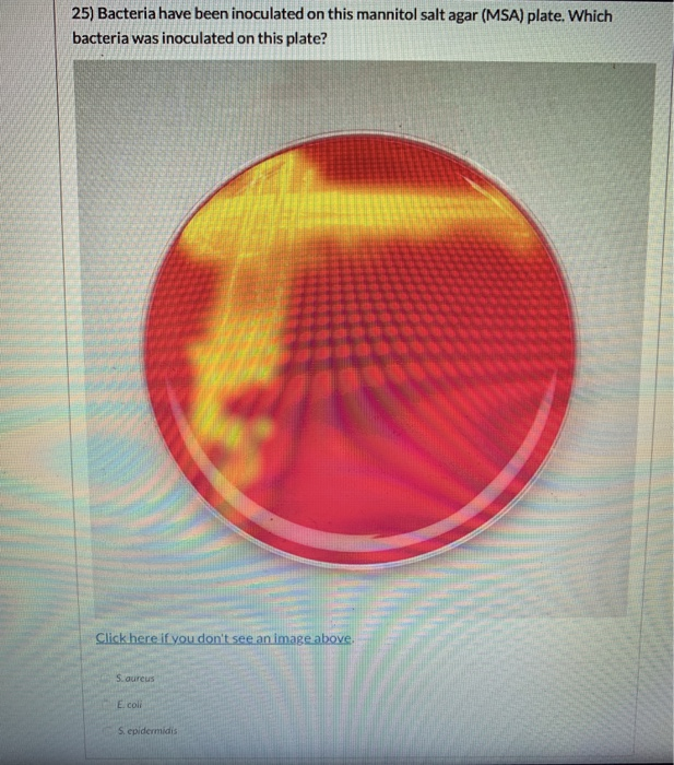
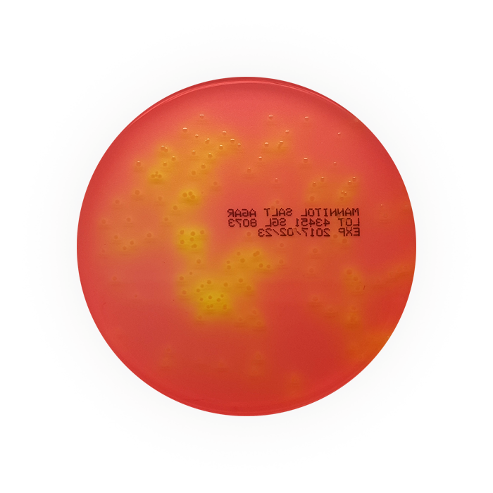
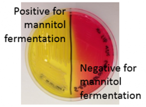
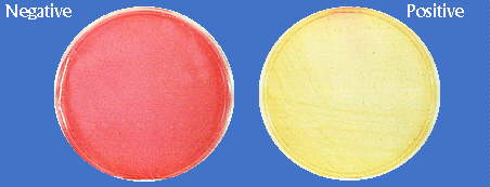
Mannitol Salt Agar (MSA) is used to determine if the bacteria is. Coli was inhibited by the high salt concentration. Is indicated by the agar media changing from red to yellow.
*Side note* MSA plate is used to test for G+ coccus. (D) Colonies of S. Mannitol salt agar (MSA) contains:.
All strains were phenotypically characterised using MSA agar plates and identified by MALDI-TOF analysis. Divide a Mannitol Salt Agar MSA plate in thirds using a Sharpie pen Label the from MICROBIOLO 24 at Northwest Vista College. Escherichia coli (Gram negative) - No growth.
A positive result would confirm Staphylococcus epidermidis as the bacterium, (4). It is recommended for the detection and enumeration of coagulase-positive Staphylococci in milk, food and other specimens and encourages the growth of a group of certain bacteria while inhibiting the growth of others. Finally, a mannitol test was performed to confirm that the bacterium is S.
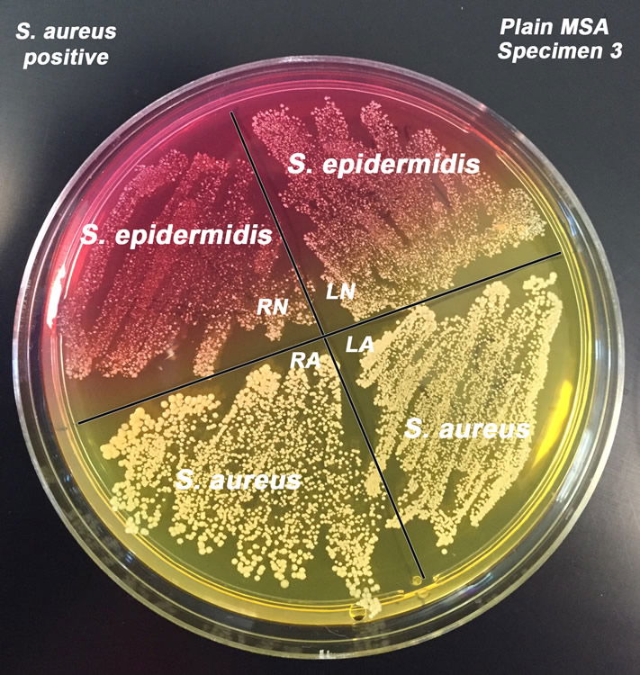
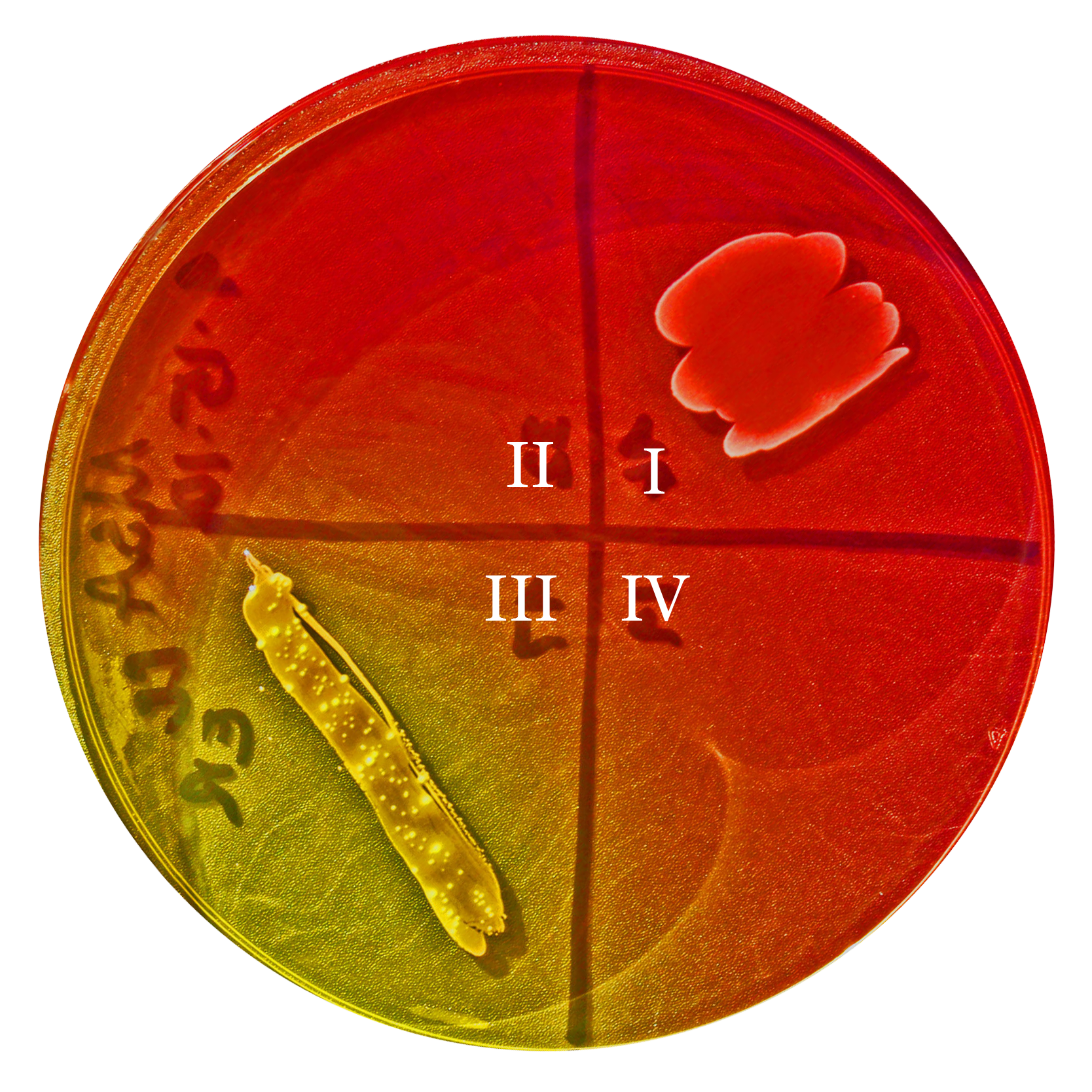
Organisms from other genera may grow, but they typically grow very weakly. (C) Colonies of S. Epidermidis, which forms colonies with red zones.
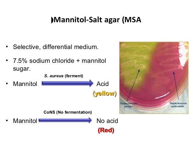
The plate contains salt and salt "loving" bacteria will grow. Some symptoms that may be common to all Staphylococcus epidermidis infections include fever, fatigue, pain or tenderness at the site of an implant, rapid breathing, rapid heartbeat and sweating. If an organism ferments mannitol, an acid will be produced turning the indicator yellow.
Staphylococcus epidermidis growing on MSA, 3 Mannitol + bacteria on left plate, Mannitol - bacteria on right plate, both plates are MSA;. Because Staphylococcus epidermidis can cause infection in such a wide variety of locations in the body, symptoms of infection are partially dependent on where the infection is. Each unknown was properly identified.
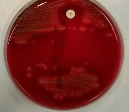
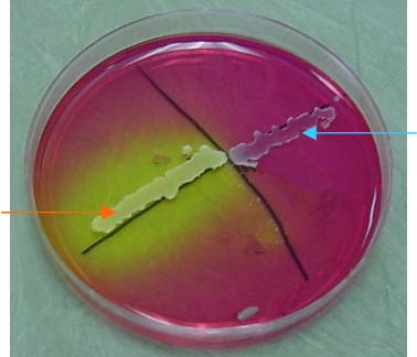
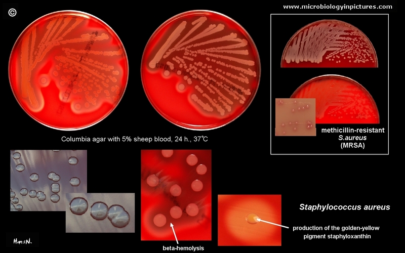
Observe hemolysis on the BAP's. It contains a high concentration (~7.5%-10%) of salt (NaCl), making it selective for gram positive bacteria. The mannitol fermenting colony (yellow) is S.
Incubate plates aerobically at 35-37ºC for 24-48 hours. 6218-07, methicillin-resistant Staphylococcus epidermidis (MRSE) 46-02, 876-09, 2490-07, 2540-07, and 4384-09, methicillin-resistant Staphylococcus haemolyticus (MRSH). A control plate of Mannitol Salt.
(1), Staphylococcus epidermidis (2) and S. What reaction does Staphylococcus epidermidis have on a MSA plate?. Staphylococcus epidermidis is a Gram-positive bacterium, and one of over 40 species belonging to the genus Staphylococcus.
Gram stains were performed on that isolated colony using the sterile technique. Selective growth of S. Streak each organism on MSA and BAP.
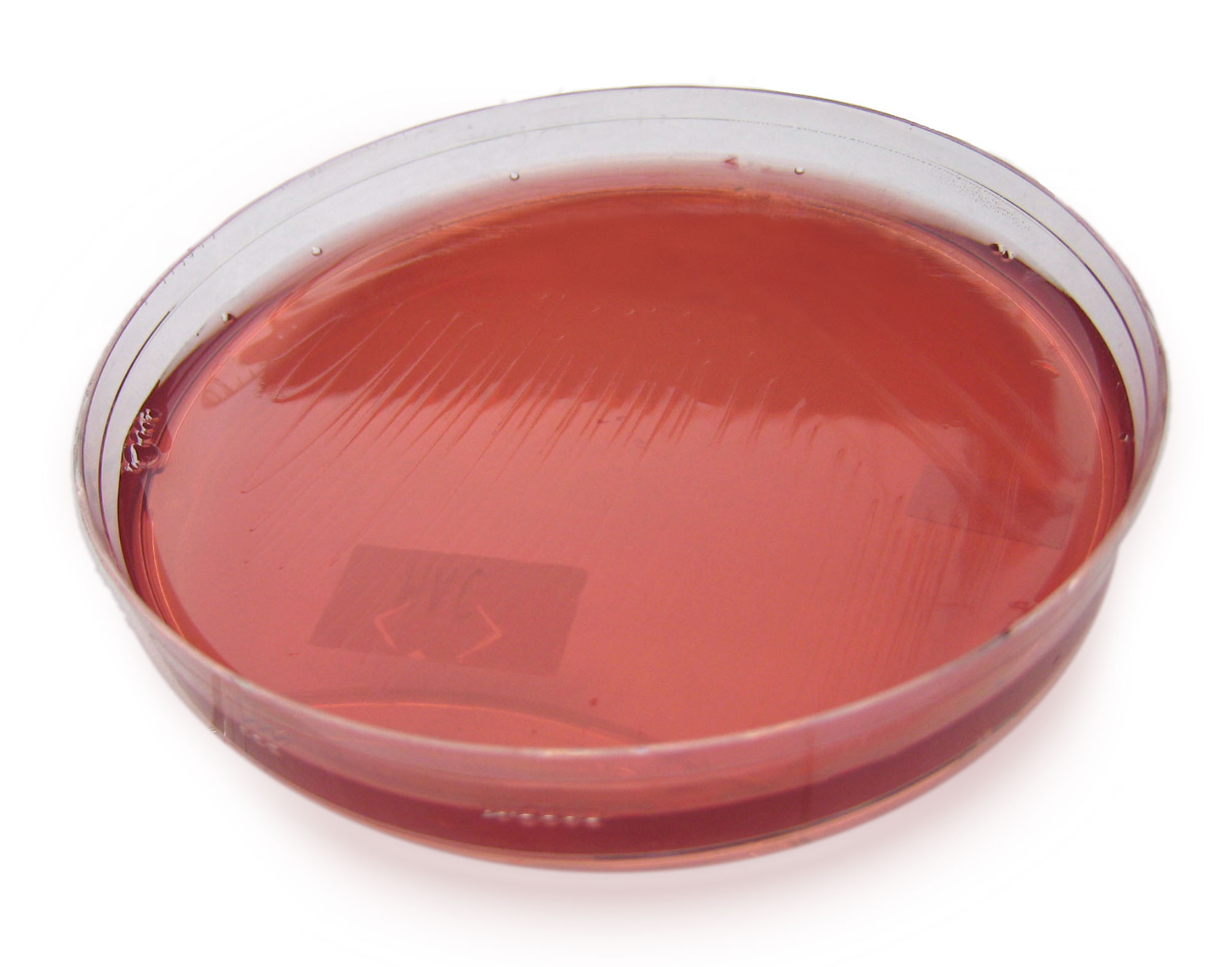
Staphylococcus epidermidis grows on MSA, but does not ferment mannitol (media remains. It encourages the growth of a group of certain bacteria while inhibiting the growth of others. This indicated that the unknown was Staphylococcus aureus or Enterococcus faecalis.
Staphylococcus epidermidis Description, Causes and Risk Factors:. Mannitol Salt Agar Mannitol Salt Agar (MSA) Plate Clinical Laboratories Science Microbiology 2 With Orwin At Mannitol Salt Agar (MSA):. The MSA plate was incubated for two days and the bacterium grew.
Psuedomonas aeruginosa (Gram negative) - no growth. Mannitol Salt Agar (MSA) is a selective and differential medium. February 13, Size:.
Composition, Uses And Colony Brightfield Microscopy Thermo Scientific Remel Mannitol Salt Agar (MSA. Mannitol Salt Agar (MSA) is used as a selective and differential medium for the isolation and identification of Staphylococcus aureus from clinical and non-clinical specimens. Does Staphylococcus grow and ferment on MSA plates?.
The bank consisted of various staphylococcal species including S. PHOTOS OF MANNITOL SALT AGAR (MSA):. Normal flora, is not a fermentor of mannitol, negative coagulase reaction.
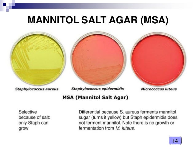
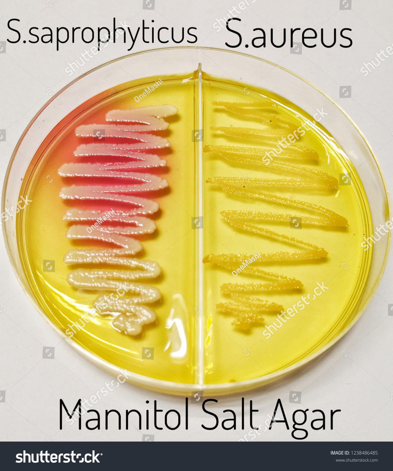
Most bacteria cannot survive in this highly saline, hypertonic environment. If one is testing among the Staphylococcus species, pathogenic Staphylococcus aureus ferments mannitol, nonpathogenic Staphylococcus species do not. Mannitol + bacteria on left, Mannitol - bacteria on right, both plated together on one MSA plate;.
Mannitol Salt is a selective bacterial growth medium because it has a very high concentration of NaCl (7.5%). Do you remember what is mannitol?. Staphylococcus epidermidis is one of the most common nosocomial (hospital acquired) infections and this is due to common use of medical devices entering the body.
Right plate non-mannitol fermenter. However, it is differential based on the ability of the organism to produce enzymes called hemolysins, which lyse red blood cells (RBC). 7.5% Which makes it selective.
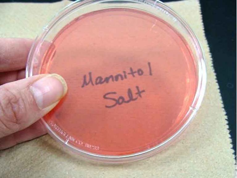
Sterile plate of Mannitol Salt agar;. It grows but does NOT ferment. Mannitol fermentation by pathogenic staphylococci, such as S.
Aureus can produce a variety of syndromes with clinical manifestations, including skin and soft tissue infections, empyema, bloodstream infections, pneumonia, osteomyelitis, septic arthritis, endocarditis, sepsis, and meningitis. Incubate enrichment per <61>. Staphylococcus epidermidis is the predominant Staphylococcus species on the human skin.
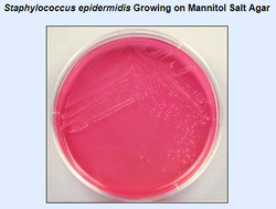
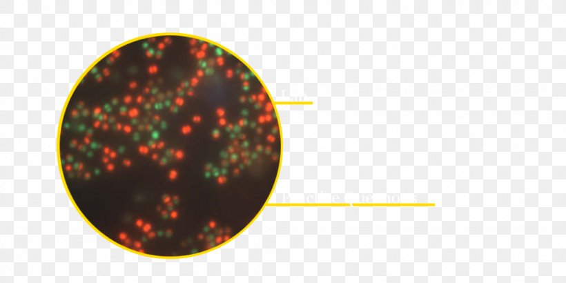
Staphylococcus epidermis growing on Mannitol Salt Agar, showing punctiform Staphylococcus epidermidis. Using a rolling motion, keep the plate in place, making sure all of the agar surface touches the testing area. 7 Staphylococcus aureus on Mannitol Salt Agar.
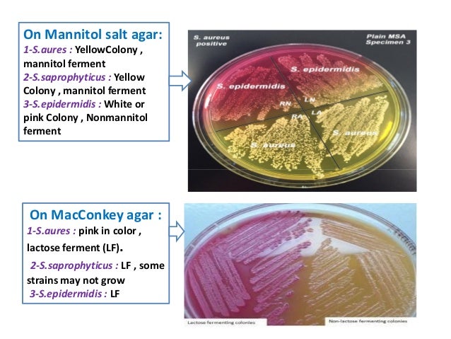
Staphylococcus aureus causes such diseases as food poisoning, meningitis, osteomyelitis and toxic shock syndrome. However, Staphylococcus epidermidis does not have the ability to ferment mannitol, because it lacks the enzyme coagulase. Aureus, while the mannitol nonfermenting colony (pink) is S.
Aureus from three discrete FDMW samples (FDMW samples 1, 2 and 3). From the MIC experiments, an acriflavine concentration of 1·5 mg l −1 was found to yield the highest reduction of S. Same plate from bottom.
Epidermidis is the species most commonly. 3) On the second of the two MSA plates, within one half of the divided plate mark label it as ‘S. Staphylococci are commonly found on the skin and in the mucous membranes of humans and other mammals.
Epidermidis is not usually pathogenic, patients with compromised immune systems are at risk of developing infection. When the skin is injured (wounds, burns, intravenous drug addicts etc), Staphylococcus epidermidis …. Sterile plate of Mannitol Salt Agar (MSA), 2.
Mannitol salt agar (MSA) is a selective, differential and indicator medium which is used to isolate and identify Staphylococcus aureus from the clinical specimen. Hominis but the first three are the most common isolates. But the genus Staphylococcus has a protective slime layerthat protects it in a harsh.
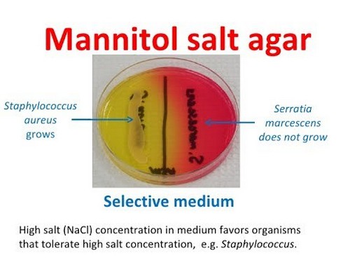
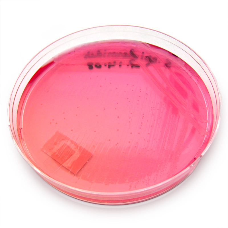
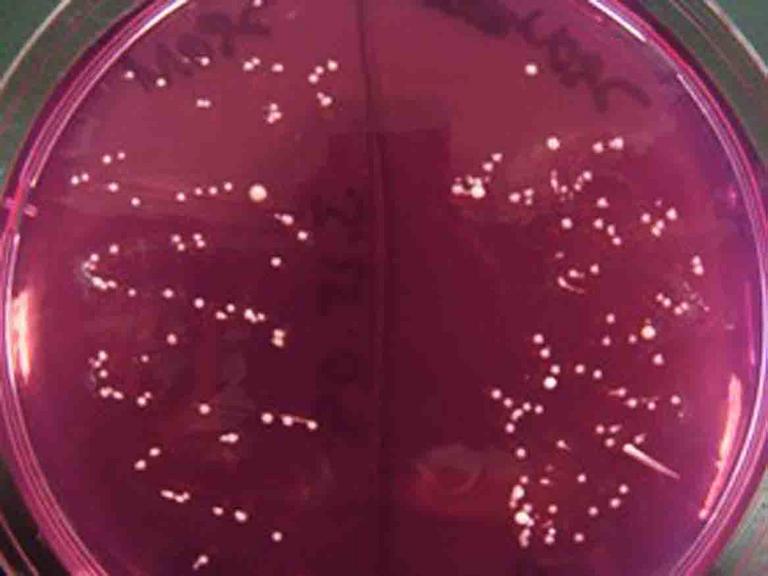
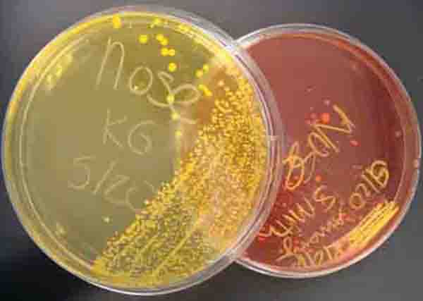
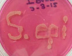
Epidermidis without greatly sacrificing S. The MSA will select for organisms such as Staphylococcus species which can live in areas of high salt concentration (plate on the left in the picture below). Mannitol nonfermenters such as Staphylococcus epidermidis will leave the MSA media unaltered (pink).
Epidermidis) is a part of a normal skin flora, and it is often attached to the upper layer of the skin (epidermis) or mucosa, without causing any symptoms (staph epidermidis carrier state). (A) Staphylococcus epidermidis cells (Gram stain). Mannitol salt agar plate (msa) Selective for gram-positive bacterium (e.g.
The high concentration of salt (7.5%) selects for members of the genus Staphylococcus, since they can tolerate high saline levels. Blood agar is a rich, non-selective medium that supports the growth of most bacteria. Epidermidis is not usually pathogenic, patients with compromised immune systems are at risk of developing infection.
After gram staining it was confirmed to be gram negative. Staphylococcus aureus is a coagulase-positive, catalase-positive, Gram-positive cocci that occur singly or in pairs, tetrads, short chains, and grape-like clusters.S. However, Staphylococcus aureus grows on MSA and fermentes giving a positive test.
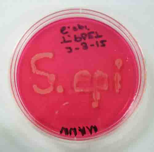
Staphylococcus epidermidis is salt-tolerant (halophilic) so it will grow on the mannitol salt agar (MSA) Plate and will produce colonies. Unknown B – Staphylococcus epidermidis DISCUSSION/CONCLUSION. What percent of NaCI does MSA plates have?.
Staphylococcus aureus (Gram positive) - growth with yellow halo. Download Image Picture detail for :. INTERPRETATION OF RESULTS.
Place the plate at 2 to 8 degree Celsius in a plastic bag in order to prevent loss of moisture. It is a facultative anaerobic bacteria. This is in contrast to Streptococcus species, whose growth is selected against by this high salt agar (plate on the right in the picture below).
Label the other half as ‘E. Mannitol Salt Agar (MSA) This type of medium is both selective and differential. Things such as catheters and heart valves can become contaminated with Staphylococcus epidermidis by contact with the skin and then act as a fomite to cause the bacterium to reach the bloodstream (2).
Non-mannitol fermentors such as S. The tests ran on the Gram-positive unknown (Staphylococcus epidermidis) were as followed:. The cocci organize into tetrads and clusters.
Aureus appear as yellow colonies with yellow zones in the media. Aureus from FDMW samples. Epidermidis cells are spherical (0.5–1.5 μm in diameter) and gram-positive.
The medium can last for a few weeks provided no abnormalities in the medium’s appearance. (1), Staphylococcus epidermidis (2) and. This culture was determined to be gram positive cocci.
Several different bacteria were plated, but only the halophiles grew. A bank of 100 CoNS strains was assembled. Staphylococcus epidermidis colonies on tryptic soy agar plate Staphylococcus epidermidis on Tryptic Soy Agar, cultivation 24 hours, 37°C Staphylococcus epidermidis is part of the normal human flora, typically the skin flora, and less commonly the mucosal flora.Although S.
Mannitol fermentors such as S. Mannitol salt agar or MSA is a commonly used selective and differential growth medium in microbiology. 2) Within one half of one divided MSA plate, label it as ‘S.
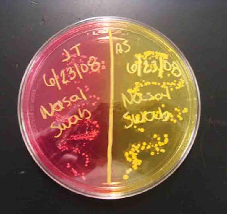
Using Figure 1 as a reference, examine your SM 110 plate for a gold colony and check your MSA. Nasal clinical samples on Mannitol Salt Agar (MSA). The second streak plate yielded only one isolated colony.
Label the other half as ‘S. Mannitol salt agar is a commonly used growth medium in microbiology. The test was performed by inoculating the mannitol broth tube with a sample from the MSA plate, to determine if the bacterium can ferment the carbohydrate mannitol as a carbon source, (3).
In the previous lab, BAP and MSA plates were inoculated with Staphylococcus and Streptococcus spp. Do not use the medium if there are any signs of abnormalities as they could indicate a possible contamination, alteration, and deterioration. The first streak plate was unsuccessful, thus the second was required.
Gently place on area to be tested. Saprophyticus and 1 S. On MSA, pathogenic Staphylococcus aureus ferments mannitol, thereby changing the colour of the medium from red to yellow.
1000px x 1000px More Galleries of Mannitol Salt Agar. What does EMB stand for?. Method of Use, Contact Plates:.
It encourages the growth of a group of certain bacteria while inhibiting the growth of others. Explain why the results on the nutrient agar plate are necessary to justify your conclusions about the mannitol salt agar plate.*. Written on MSA with the bacteria Staphylococcus epidermidis, a halophilic, non-mannitol fermenter;.
(A) Staphylococcus aureus, (B) Staphylococcus epidermidis, and (C) Escherichia coli streaked on a mannitol salt agar plate. Epidermidis incubated on agar plate. Observe for salt tolerance and mannitol fermentation on the MSA plates.
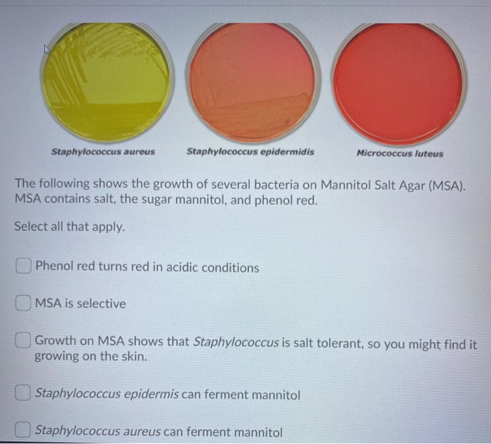
Aureus.MSA supplemented with 1·5 mg l −1 acriflavine was tested for quantifying indigenous S. MSA also contains the sugar mannitol and the pH indicator phenol red. Incubate the plates at 37°C for 48 hours.
MSA plates were also incubated at 34 C according to the manufacturer’s rec-ommendations. MSA is a Differential Medium because of the presence of the sugar mannitol and the pH indicator Phenol Red.
Mannitol Salt Agar Plate Test Composition Preparation Uses Laboratoryinfo Com

Mannitol Salt Agar Wikipedia

Got Science Notanotherscienceblog Staphylococcus Aureus On

Mannitol Salt Agar Medical Laboratory Scientists Medical Laboratory Scientist Medical Laboratory Science Microbiology Lab
Cultivation Media For Bacteria
Solved Staphylococcus Aureus Staphylococcus Epidermidis M Chegg Com

Mannitol Salt Agar Msa Test Superfarmer S Weblog
Mannitol Salt Bacterial Growth Medium Msa Page 2
Sketchy Micro Bacteria Staph Aureus The Golden Staff Of Moses Flashcards Memorang
Mannitol Salt Bacterial Growth Medium Msa Page 2

Mannitol Salt Agar Medical Laboratory Scientist Medical Laboratory Science Medical Laboratory
Solved 4 3 Mannitol Salt Agar Observations And Interpret Chegg Com

Staphylococcus Mannitol Salt Agar Plate Medical Laboratories

A Staphylococcus Aureus And Staphylococcus Epidermidis Grown On Download Scientific Diagram

Lab Manual Superfarmer S Weblog

Selective And Differential Media Ppt Video Online Download

Mannitol Salt Agar Staphylococcus Aureus Msa Youtube
I Ve Attached Lab Worksheets And Images Phenylet Chegg Com
Www Jfmed Uniba Sk Fileadmin Jlf Pracoviska Ustav Mikrobiologie A Imunologie Vla Staphylococci Pdf

Mannitol Salt Agar Chapman Medium Staphylococcus Aureus And Staphylococcus Epidermidis Colonies On Mannitol Salt Agar Plate

Mannitol Salt Agar Msa For The Identification Of Staphylococcus Aureus And Epidermidis
Mannitol Salt Agar Msa Images Stock Photos Vectors Shutterstock
Solved 25 Bacteria Have Been Inoculated On This Mannitol Chegg Com

Fermentation Of Mannitol Salt Agar By S Aureus Formation Of Yellow Download Scientific Diagram
L13 Medical Microbiology Laboratory Staphylococcus Spp

Staphylococcus Aureus And Staphylococcus Epidermidis On Mannitol Salt Agar
Article Fulle Text
1

Mannitol Salt Agar Msa Composition Uses And Colony Characteristics Learn Microbiology Online

Pin On Tasha Sturm Everything Microbiology

Mannitol Salt Agar Microbiology Food And Drug Reference
Staph Bacteria From First Breath Answers In Genesis
Mannitol Salt Agar 90mm Plate Southern Group Laboratory
Q Tbn 3aand9gcq0fb7kqcd3bewi3vuqqhupwlp4bqj Qjkilrelsirym2lkqaox Usqp Cau

Lab 5 Integumentary System Bacteriology And Identification Ppt Download
Staphylococcus
Mannitol Salt Agar Plate Test Composition Preparation Uses Laboratoryinfo Com

Staphylococcus Epidermidis On Agar Plate With Mannitol Salt Agar Chapman Agar Growth Of S Epidermidis In Petri Dish On Chapman Agar Medium Appearance And Morphology Of Staphylococcus Epidermidis And S Aureus Colonies On Mannitol
Staphylococcus Aureus Staphylococcus Saprophyticus On Mannitol Stock Photo Edit Now

Microbiology Notes And Updates Growth Of Staphylococcus Aureus Yellow And Staphylococcus Epidermidis Red On Mannitol Salt Agar Plate Image By Matthew Donadu Facebook

Isolation And Identification Of Staphylococci Ppt Video Online Download
Mannitol Salt Agar Msa Test Scientist Cindy
Staphylococci Prac Microbiology

Growth On Mannitol Salt Agar Media Showing Presence Of Yellow Halo Download Scientific Diagram
Welcome To Microbugz Mannitol Salt Agar
Metabolism Physiology And Growth Characteristics Of Cocci Microbiology A Laboratory Experience

Mannitol Salt Agar Microbiology Food And Drug Reference

A Staphylococcus Aureus And B Staphylococcus Epidermidis On Download Scientific Diagram
Cultivation Media For Bacteria
Mannitol Salt Agar Microbiology Images Photographs From Science Prof Online
Mannitol Salt Agar Staphylococcus Aureus Staphylococcus Epidermidis Agar Plate Halophile Png 1024x512px Mannitol Salt Agar Agar
Solved Stoph Epidermidis Mannitol Salt Agar Plate Resul Chegg Com
Experiment 8c Lab08 Virtual Edge Molb 21 College Of Agriculture And Natural Sciences

Staphylococcus Aureus On Mannitol Salt Phenol Red Agar Stock Photo Download Image Now Istock

Mannitol Salt Agar Li 30ml Application Isolation And Presumptive Identification Of Staphylococcus Aureus In Non Sterile

Isolation Identification Of Staphylococci Ppt Video Online Download

Mannitol Salt Agar Msa Diagram Quizlet

Staphylococcus Aureus And Staphylococcus Epidermidis On Mannitol Salt Agar Mannitol Salt Agar Composition Positive And Negative Result On Mannitol Salt Agar

Staphylococcus Epidermidis Under Microscope Microscopy Of Gram Positive Cocci Morphology And Microscopic Appearance Of Staphylococcus Epidermidis S Epidermidis Gram Stain And Colony Morphology On Agar Clinical Significance
Microbiology Lab Molb 2210
Mannitol Salt Bacterial Growth Medium Msa Page 2

Asmscience Mannitol Salt Agar
Untitled Document

Pink Colonies Of Staphylococcus Epidermidis Isolate Grow On Mannitol Download Scientific Diagram

Media Examples With Pics Flashcards Quizlet

Typical Golden Yellow Colonies Of Staphylococcus Aureus On Mannitol Download Scientific Diagram
Mannitol Salt Agar Microbiology Images Photographs From Science Prof Online
Microbiology Lab Molb 2210

Solved With The Pictures Of The Tests Mannitol Salt Mac Chegg Com

The Belly Button Can Act As A Reservoir For Staphylococcus Aureus Cleaning The Area With Chlorhexidine Can Reduce And Possibly Eliminate Its Presence Staphacne

Selective Growth Of Staphylococcus Aureus From Flushed Dairy Manure Wastewater Using Acriflavine Supplemented Mannitol Salt Agar Davis 06 Letters In Applied Microbiology Wiley Online Library
Http Www vmc Org Assets Site 18 Files Case Study Burnham staph pseudintermedius student materials Pdf

Mannitol Salt Agar Msa Composition Uses And Colony Characteristics Learn Microbiology Online
1
Q Tbn 3aand9gctgrmjg8acjrbxbniknzy1qewngtoobg8cixbwsv Ok9ny02ti0 Usqp Cau
Mannitol Salt Agar Microbiology Images Photographs From Science Prof Online
What Is Mannitol Salt Agar Biology Wise
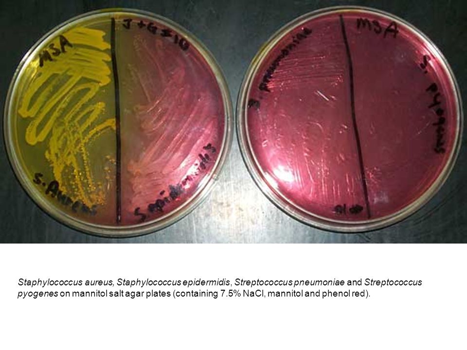
Staphylococcus Aureus Staphylococcus Epidermidis Streptococcus Pneumoniae And Streptococcus Pyogenes On Mannitol Salt Agar Plates Containing Ppt Download

Growth Of Staphylococcus Aureus On Mannitol Salt Agar Download Scientific Diagram

Mannitol Salt Agar Plate With Oxacillin Showing Methicillin Resistant Download Scientific Diagram

Mannitol Salt Agar Msa Results Theory Youtube
Mannitol Salt Agar
Pht 313 Lab 1 Staphylococci Ppt Video Online Download
Mannitol Salt Agar Msa Plate
_(1).jpg?bwg=1562148427)
Micr350 Streaked Images

How To Interpret Mannitol Salt Agar Bacterial Growth Medium Youtube
Mannitol Salt Agar For The Isolation Of Staphylococcus Aureus

Mannitol Salt Agar Test Msa Flashcards Quizlet

Staphylococcus Aureus On Combined Mannitol Salt Agar And Vogel Johnson Agar Microbiology Pictures Microbe Notes

Product Catalog
Mannitol Salt Agar Msa Plate

Study Selective Differential Media Flashcards Quizlet
Mannitol Salt Bacterial Growth Medium Msa Page 2

Staphylococcus Aureus On Mannitol Salt Phenol Red Agar Stock Photo Download Image Now Istock
Staphylococcus Aureus Colony Morphology And Microscopic Appearance Basic Characteristic And Tests For Identification Of S Aureus Bacteria Images Of Staphylococcus Aureus Antibiotic Treatment Of Staphylococcal Infections

Mannitol Salt Agar Msa For The Identification Of Staphylococcus Aureus And Epidermidis

Biol 230 Lab Manual Staphylococcus Epidermidis On Mannitol Salt Agar


